Before an MRI examination, due to the strong magnetic field that prevails here, all magnetic / metallic objects that you wear on your body must be taken off for safety reasons. These include E.g. keys, chip cards, money, watches, glasses, hearing aids, jewelry, removable dentures.
If you have metallic materials / implants in your body, complications can arise due to heating / damage to the material. For safety reasons, an examination with magnetic resonance tomography is unfortunately not possible in the following cases:
Pacemaker / defibrillator
Neurostimulators
In all of these cases, you must inform us prior to the investigation. Our experienced team of doctors is happy to assist you with any questions or doubts.
For claustrophobia:
It is not uncommon for patients to have claustrophobia problems in the examination device. Please do not hesitate to point this out to our practice team. We are particularly sensitive to patients with fears and find a common solution so that you can relax optimally.
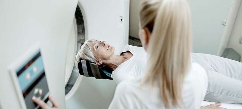
The examination usually takes between 15-30 minutes and takes place lying down in a tube that is open to the foot and the head and is adequately ventilated and illuminated. You should lie as still as possible during the examination. There is both eye contact with our staff and voice contact through an audio system. In addition to this, every patient receives an emergency button in their hand that they can trigger at any time. Since the examinations can cause louder knocking noises, wear sound-absorbing headphones. We monitor you throughout the entire investigation and are always available for you.
In certain cases it may be necessary to give you a contrast medium to enhance the tissue contrast during the examination. The contrast media are usually very well tolerated and only rarely cause side effects or allergic reactions.
After the examination, the radiologist will discuss the result with you if you wish and advise you in detail on any further steps.